


Jaundice: Causes, Symptoms & When to See a Doctor
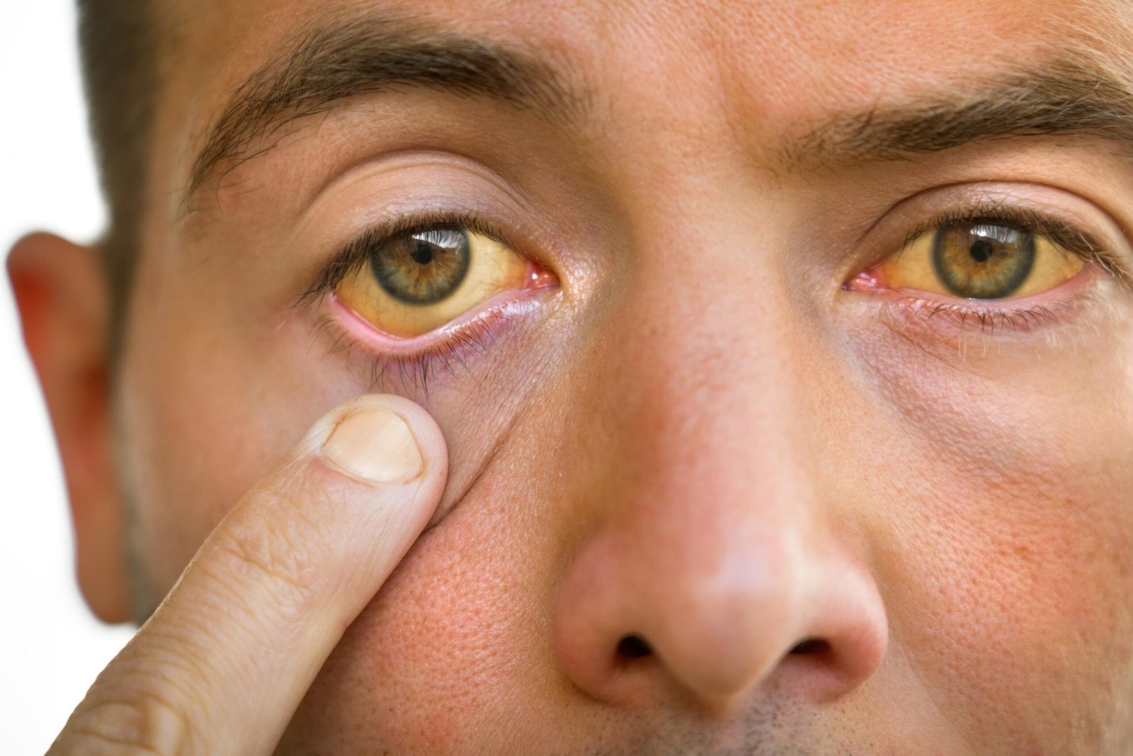
Jaundice is a visible sign of an underlying medical condition caused by a buildup of bilirubin in the bloodstream producing the characteristic yellow color in the skin, whites of the eyes, and mucous membranes. It is not a disease in itself but rather a clinical sign that points to a disruption in the way the body produces, processes, or eliminates bilirubin. Jaundice symptoms can appear in people of all ages from newborn babies in their first days of life to adults with liver disease, bile duct obstruction, or blood disorders. Hepatologists, gastroenterologists, neonatologists, and specialists at SRM Global Hospitals in Chennai evaluate jaundice comprehensively identifying the underlying cause and initiating targeted treatment to prevent serious complications.
What Is Jaundice?
Jaundice develops when bilirubin a yellow pigment produced by the normal breakdown of red blood cells accumulates in the blood faster than the liver can process and eliminate it.
Under normal circumstances, the liver processes bilirubin and excretes it into bile, which flows through the bile ducts into the digestive tract and is eliminated through bowel movements. When this process is disrupted whether due to excessive red blood cell breakdown, liver dysfunction, or obstruction of the bile ducts excess bilirubin accumulates in the blood and deposits in the skin, whites of the eyes, and mucous membranes, producing the yellow color that characterises jaundice.
Bilirubin exists in two main forms in the body:
Unconjugated bilirubin fat-soluble bilirubin not yet processed by the liver. High levels of unconjugated bilirubin are particularly dangerous in newborn babies, as unconjugated bilirubin can cross the blood brain barrier and cause bilirubin encephalopathy a serious neurological complication.
Conjugated bilirubin water-soluble bilirubin that has been processed by the liver and is ready for excretion. Elevated conjugated bilirubin typically indicates liver disease or obstruction of the bile ducts.
Understanding whether the excess bilirubin is unconjugated or conjugated is the first step toward identifying the underlying cause of jaundice and selecting appropriate treatment.
Jaundice Symptoms: What Does Jaundice Look Like?
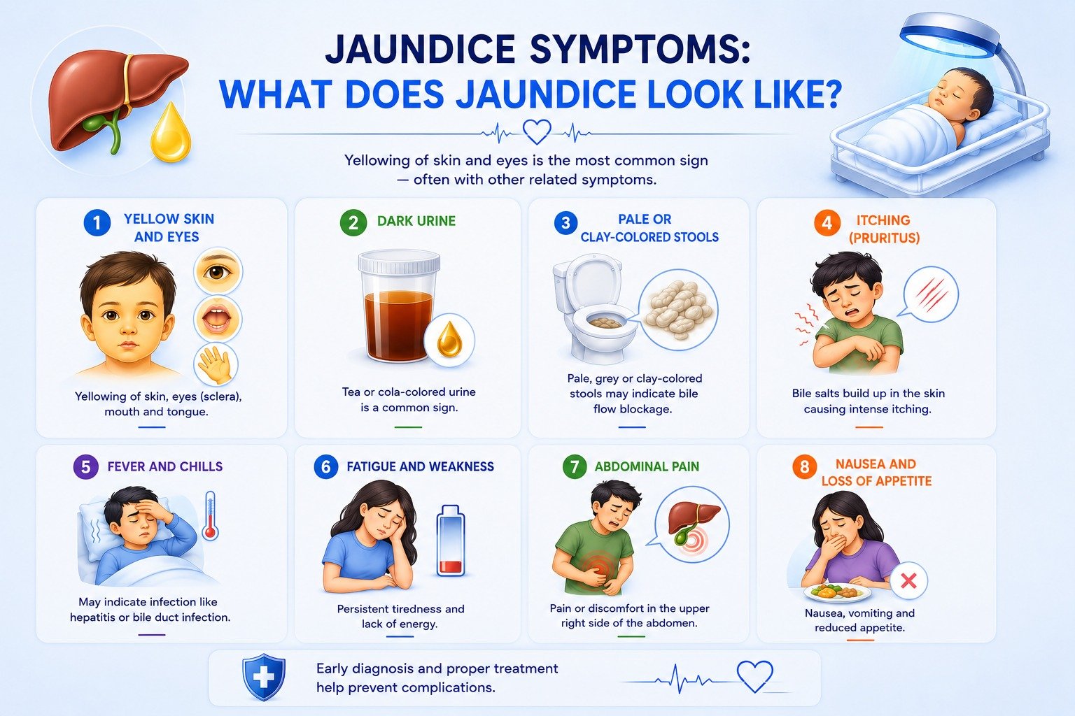
Jaundice symptoms vary depending on the underlying cause, the severity of bilirubin accumulation, and the patient’s age. The most universally recognised jaundice symptom is the yellow color of the skin and eyes but jaundice is almost always accompanied by other symptoms that reflect the specific underlying condition driving bilirubin accumulation.
The Yellow Color of Skin and Eyes
The main sign of jaundice is the yellowing of the skin, which is most visible in natural light particularly on the face first. As bilirubin levels increase, the yellow color may spread from the face to the chest, abdomen, arms, and legs in a predictable cephalocaudal progression. The whites of the eyes (sclera) are often the earliest and most sensitive site of visible jaundice even mild jaundice may produce visible yellowing of the sclera before the skin colour changes.
The mucous membranes including the inner lining of the mouth and the underside of the tongue also take on a yellow color in jaundice.
In darker-skinned individuals, jaundice may be harder to detect in the skin, but the yellowing can still be clearly observed in the whites of the eyes and under the tongue.
Dark Urine
Dark urine tea-coloured or cola-coloured urine is one of the most important associated jaundice symptoms. Dark urine occurs because excess conjugated bilirubin is excreted through the kidneys rather than through bile, staining the urine a deep yellow or brown colour. Dark urine and pale stools are commonly associated with jaundice, indicating potential liver dysfunction or bile obstruction.
Pale or Clay-Coloured Stools
Pale or clay-coloured stools indicate that bile is blocked from reaching the digestive tract which is strongly associated with jaundice caused by obstruction of the bile ducts. Normally, bile pigments give stools their characteristic brown colour. When the bile ducts are obstructed, bile cannot reach the intestine and stools become pale, grey, or clay-coloured.
Itching (Pruritus)
Severe itching called pruritus can occur due to the accumulation of bile salts in the skin associated with jaundice. Pruritus is particularly prominent in obstructive jaundice, where the bile ducts are blocked and bile salts back up into the bloodstream. The itching can be intense and significantly affects quality of life.
Fever and Chills
Fever and chills accompanying jaundice may indicate an active infection such as viral hepatitis or a bile duct infection (cholangitis). The combination of jaundice, fever, and right upper abdominal pain is known as Charcot’s triad and is a classic presentation of cholangitis requiring urgent medical attention.
Fatigue and Weakness
Persistent fatigue and general weakness are common in jaundice, reflecting the metabolic burden placed on the body by liver dysfunction or haemolytic anaemia. Patients with liver disease-related jaundice frequently report debilitating fatigue as one of their most troublesome symptoms.
Abdominal Pain
Right upper quadrant abdominal pain or generalised abdominal discomfort accompanies jaundice in conditions such as gallstones blocking the bile ducts, liver injury, or liver disease. Severe, colicky right upper abdominal pain that radiates to the back or shoulder with jaundice suggests gallstone-related bile duct obstruction.
Nausea and Loss of Appetite
Nausea, vomiting, and reduced appetite are common in jaundice associated with liver disease particularly viral hepatitis. These symptoms reflect hepatic dysfunction and the body’s inflammatory response to liver injury.
When Jaundice Symptoms Require Immediate Medical Attention
Seek emergency care at SRM Global Hospitals immediately if jaundice is accompanied by:
Emergency Symptom | What It May Indicate |
|---|---|
Severe abdominal pain with jaundice | Acute bile duct obstruction, liver failure, or pancreatitis |
High fever, rigors, and jaundice | Cholangitis (bile duct infection) a surgical emergency |
Confusion or altered consciousness | Hepatic encephalopathy from acute liver failure |
Rapid deterioration in jaundice severity | Acute liver failure requiring intensive care |
Jaundice in a newborn baby within 24 hours of birth | Pathological neonatal jaundice immediate assessment required |
Causes of Jaundice
Jaundice can be classified into three phases based on where in the bilirubin processing pathway the disruption occurs: pre-hepatic, hepatic, and post-hepatic. Each phase has distinct causes of jaundice, produces a different pattern of bilirubin accumulation, and requires a different treatment approach.
Pre-Hepatic Jaundice (Before the Liver)
Pre-hepatic jaundice occurs when red blood cells are destroyed faster than the liver can process the resulting bilirubin. This excessive red blood cell breakdown called haemolysis floods the liver with more unconjugated bilirubin than it can handle, causing elevated unconjugated bilirubin in the bloodstream.
Causes of pre-hepatic jaundice include:
Haemolytic anaemia — autoimmune haemolytic anaemia, sickle cell disease, thalassaemia, and hereditary spherocytosis all cause accelerated destruction of red blood cells
Blood group incompatibilities — ABO and Rh incompatibilities between mother and baby cause immune-mediated haemolysis, particularly in newborn infants
Malaria — the malaria parasite destroys red blood cells directly, producing haemolytic jaundice
G6PD deficiency — a hereditary red blood cell enzyme defect that causes episodic haemolysis and jaundice when triggered by certain medications or foods
Sepsis — severe infection causes haemolytic disease and jaundice through multiple mechanisms
Hepatic Jaundice (Within the Liver)
Hepatic jaundice occurs when the liver itself is diseased or damaged and cannot process bilirubin normally regardless of whether the production rate of bilirubin is normal or elevated.
Causes of hepatic jaundice include:
Viral hepatitis (Hepatitis A, B, C, D, E) — viral infection of liver cells impairs the liver’s ability to process bilirubin, causing both unconjugated and conjugated bilirubin to accumulate
Alcoholic liver disease — chronic excessive alcohol consumption causes liver injury, hepatitis, fibrosis, and ultimately cirrhosis — all of which impair bilirubin processing
Non-alcoholic fatty liver disease (NAFLD) and NASH — fat accumulation in liver cells impairs liver function and can progress to cirrhosis
Cirrhosis — end-stage liver disease from any cause where extensive scarring replaces functional liver tissue, severely impairing the liver’s ability to process bilirubin
Drug-induced liver injury — certain medications including paracetamol overdose, statins, antitubercular drugs, and herbal preparations can cause liver injury and jaundice
Autoimmune hepatitis — the immune system attacks liver cells, causing progressive liver injury
Liver tumours — primary liver cancer (hepatocellular carcinoma) or metastatic disease replacing functional liver tissue
Post-Hepatic Jaundice (After the Liver — Obstructive Jaundice)
Post-hepatic jaundice also called obstructive jaundice or cholestatic jaundice occurs when the bile ducts are blocked, preventing processed conjugated bilirubin from reaching the digestive tract. Blocked bile backs up into the liver and bloodstream, causing elevated conjugated bilirubin and the characteristic dark urine and pale stools.
Causes of post-hepatic (obstructive) jaundice include:
Gallstones in the bile ducts (choledocholithiasis) — the most common cause of obstructive jaundice in adults; gallstones migrate from the gallbladder and block the common bile duct
Biliary atresia — a serious congenital condition in which the bile ducts are absent or damaged from birth, causing progressive jaundice in newborn babies. Biliary atresia requires early surgical intervention and is one of the most important causes of jaundice in infants.
Pancreatic cancer — a tumour at the head of the pancreas compresses the bile duct, causing progressive painless jaundice — often one of the earliest warning signs
Cholangiocarcinoma — cancer of the bile ducts
Primary sclerosing cholangitis (PSC) — inflammatory narrowing of the bile ducts, commonly associated with inflammatory bowel disease
Primary biliary cholangitis (PBC) — autoimmune destruction of small bile ducts within the liver
Jaundice in Newborns (Neonatal Jaundice)
Neonatal jaundice jaundice in newborn babies is one of the most common medical conditions encountered in the newborn nursery. Newborn jaundice requires careful assessment and monitoring because elevated bilirubin levels particularly unconjugated bilirubin can cross the blood brain barrier and cause bilirubin encephalopathy, leading to serious neurologic complications if left untreated.
Why Do Babies Develop Jaundice?
Newborn babies develop jaundice far more readily than adults because of several physiological factors specific to neonatal bilirubin metabolism:
Newborn babies have a higher concentration of red blood cells than adults, and these fetal red blood cells have a shorter lifespan producing more bilirubin from their breakdown
A newborn baby’s liver is not yet fully mature and has a limited capacity to process bilirubin efficiently
The gut of a newborn infant has lower bacterial colonisation, which means less bilirubin is converted in the intestine and more is reabsorbed back into the bloodstream (enterohepatic circulation)
These combined factors mean that most newborn babies experience some degree of bilirubin level elevation in the first week of life.
Types of Neonatal Jaundice
Physiological Jaundice
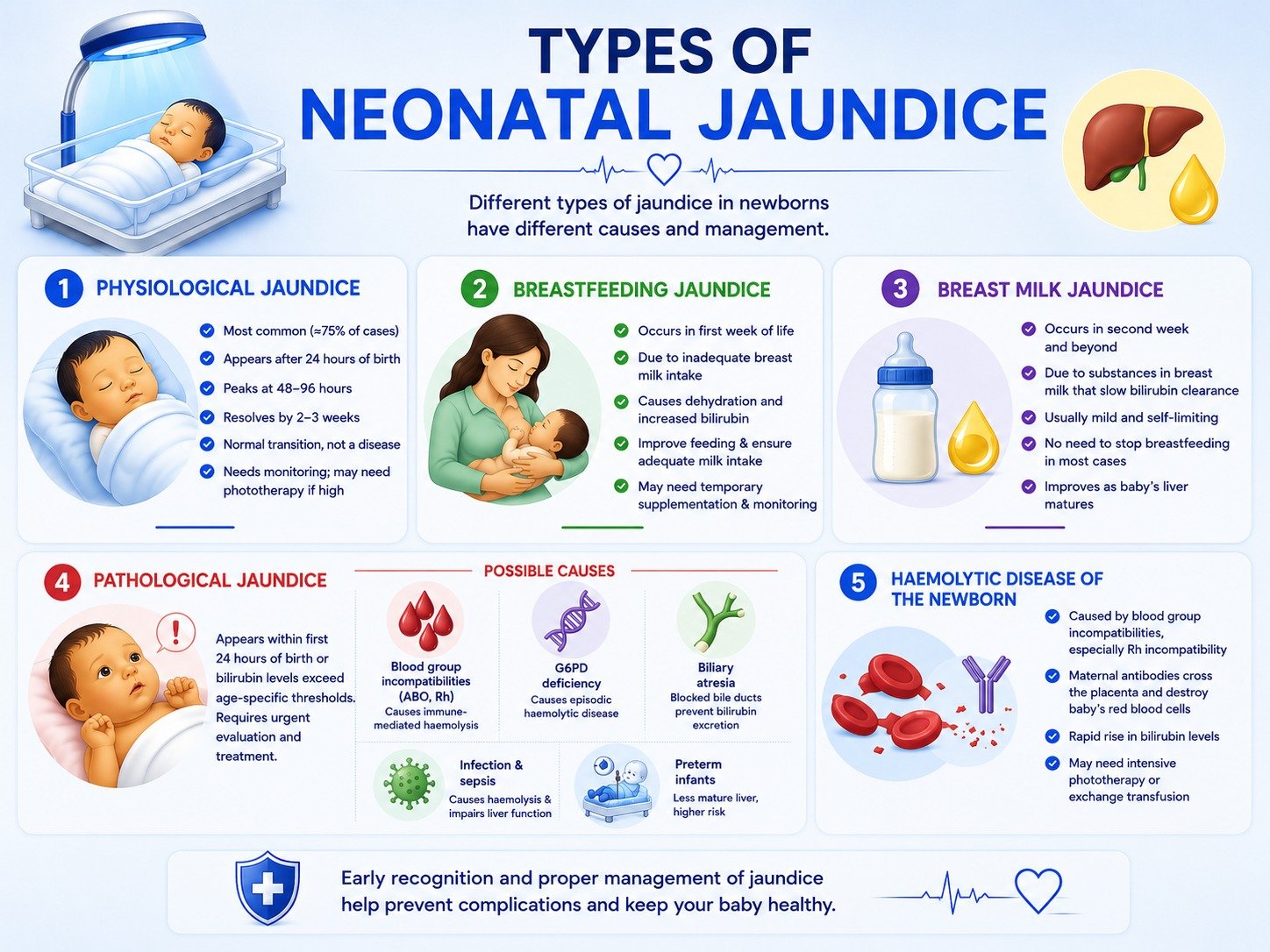
Physiological jaundice is the most common type of jaundice in newborns, accounting for approximately 75% of cases of neonatal hyperbilirubinemia. It typically appears 24 hours after birth, peaks at 48 to 96 hours, and resolves by 2 to 3 weeks in most newborn babies. Physiological jaundice is caused by the normal transition from fetal to neonatal bilirubin metabolism and does not indicate underlying disease. However, even physiological jaundice must be monitored, as bilirubin levels can occasionally rise to levels requiring phototherapy treatment.
Breastfeeding Jaundice
Breastfeeding jaundice occurs when a newborn does not receive enough breast milk leading to dehydration and increased bilirubin levels. It is more common in the first week of life and is related to inadequate feeding rather than any problem with the breast milk itself. Supporting the mother to establish effective breastfeeding and ensuring adequate breast milk intake is the primary management strategy. Breastfed infants with significant breastfeeding jaundice may require temporary supplementation and close bilirubin monitoring.
Breast Milk Jaundice
Breast milk jaundice is a distinct condition from breastfeeding jaundice. It occurs in breastfed infants in the second week of life and beyond, caused by substances in breast milk that inhibit bilirubin clearance in the baby’s liver. Breast milk jaundice is typically mild jaundice, self-limiting, and does not require stopping breastfeeding in most cases. The baby’s bilirubin level gradually decreases as the infant’s liver matures, even while breastfeeding continues.
Pathological Jaundice
Pathological jaundice also called pathological unconjugated hyperbilirubinemia occurs within the first 24 hours after birth and is characterised by a total serum bilirubin level that exceeds age-specific thresholds, indicating a need for further evaluation. Pathological jaundice requires urgent investigation to identify the underlying cause and initiate treatment before bilirubin levels rise to levels that risk bilirubin toxicity.
Pathological causes of neonatal jaundice include:
Blood group incompatibilities — ABO and Rh incompatibilities between the mother and baby cause immune-mediated haemolysis in the baby’s blood, releasing large amounts of bilirubin from the destroyed baby’s red blood cells
G6PD deficiency — causes episodic haemolytic disease in the newborn infant
Biliary atresia — obstructed bile ducts in the baby’s liver prevent bilirubin excretion
Infection and sepsis — neonatal sepsis causes haemolytic disease and impairs the baby’s liver function simultaneously
Preterm infants — premature infants have a less mature liver and a higher risk of neonatal hyperbilirubinemia than term newborn babies
Haemolytic Disease of the Newborn
Haemolytic disease caused by blood group incompatibilities particularly Rh incompatibility causes rapid destruction of the baby’s red blood cells by maternal antibodies that have crossed the placenta. The baby’s blood is flooded with bilirubin from haemolysis, causing rapidly rising serum bilirubin levels that can quickly reach dangerous concentrations requiring intensive phototherapy or exchange transfusion.
Jaundice Symptoms in Newborns
The main sign of jaundice in newborns is the yellowing of the baby’s skin, which is most visible in natural light particularly on the face first. As bilirubin levels increase, the yellow color spreads from the face to the chest, abdomen, arms, and legs. The whites of the baby’s eyes and mucous membranes also develop yellow color.
Additional signs of severe jaundice in newborn babies include:
High-pitched crying or unusual irritability
Poor feeding or refusing breast milk or formula
Lethargy or difficulty waking the newborn infant
Arching of the back (opisthotonus) a sign of bilirubin encephalopathy
Abnormal muscle tone either too stiff or too floppy
Neonatal jaundice is harder to detect in darker-skinned newborn babies but the yellowing can still be observed in the whites of the eyes and under the tongue. The baby’s skin should be assessed in natural light for the most accurate visual assessment.
Assessment and Monitoring of Newborn Jaundice
The American Academy of Pediatrics (AAP) recommends that all newborns be assessed for jaundice every 12 hours from the time of birth until discharge. Bilirubin levels should be measured between 24 and 48 hours after birth.
Methods for measuring bilirubin levels in newborn babies include:
Transcutaneous bilirubin measurement — a non-invasive device pressed against the infant’s skin measures bilirubin level through the skin. Transcutaneous bilirubin measurement is used for initial screening but requires confirmation with a serum bilirubin test if levels are elevated.
Total serum bilirubin (TSB) — a blood test measuring the total amount of bilirubin in the baby’s blood. Total serum bilirubin is the definitive measurement used to guide treatment decisions.
Bilirubin albumin ratio — the ratio of bilirubin to serum albumin in the blood reflects the amount of free unconjugated bilirubin capable of crossing the blood brain barrier. A high bilirubin albumin ratio indicates elevated neurotoxicity risk.
The baby’s bilirubin level is plotted on a nomogram against baby’s age and gestational age to determine whether phototherapy is required and to assess neurotoxicity risk factors.
How Is Jaundice Diagnosed?
Medical evaluations to confirm jaundice and identify the underlying cause include physical exams, bilirubin and blood tests, and liver function tests. The diagnostic approach at SRM Global Hospitals is systematic starting with clinical assessment and escalating to imaging and liver biopsy where required.
Clinical Assessment
The hepatologist or specialist examines the patient for yellow color in the skin, whites of the eyes, and mucous membranes as well as for associated signs including dark urine, pale stools, abdominal tenderness, liver enlargement, and signs of chronic liver disease.
Blood Tests
Blood tests are the cornerstone of jaundice investigation:
Serum bilirubin — total serum bilirubin, along with direct (conjugated) and indirect (unconjugated) bilirubin fractions, to classify the type of jaundice
Liver function tests (LFTs) — measuring ALT, AST, ALP, GGT, and albumin to assess liver cell damage and bile duct obstruction
Full blood count — to detect haemolytic anaemia and assess red blood cells for evidence of haemolysis
Blood group and Coombs test — particularly important in newborn babies to identify blood group incompatibilities and immune-mediated haemolytic disease
Viral hepatitis serology — hepatitis A, B, C, D, and E antibodies and antigens
Coagulation studies — prothrombin time reflects the severity of liver dysfunction
Imaging Studies
Ultrasound — the first-line imaging study for jaundice, used to detect gallstones, dilated bile ducts, liver disease, and biliary atresia in babies
MRCP (Magnetic Resonance Cholangiopancreatography) — detailed non-invasive imaging of the bile ducts to identify obstruction without requiring a procedure
CT scan — used when ultrasound findings are inconclusive or when malignancy of the liver or pancreas is suspected
ERCP (Endoscopic Retrograde Cholangiopancreatography) — both diagnostic and therapeutic for bile duct obstruction; allows stone removal or stent placement during the same procedure
Liver Biopsy
A liver biopsy a small sample of liver tissue obtained through the skin may be required when non-invasive investigations have not identified the underlying cause, when autoimmune hepatitis or specific liver diseases are suspected, or when the degree of liver injury and fibrosis needs accurate staging. Liver biopsy is performed under ultrasound guidance at SRM Global Hospitals by experienced hepatologists.
Treatment for Jaundice
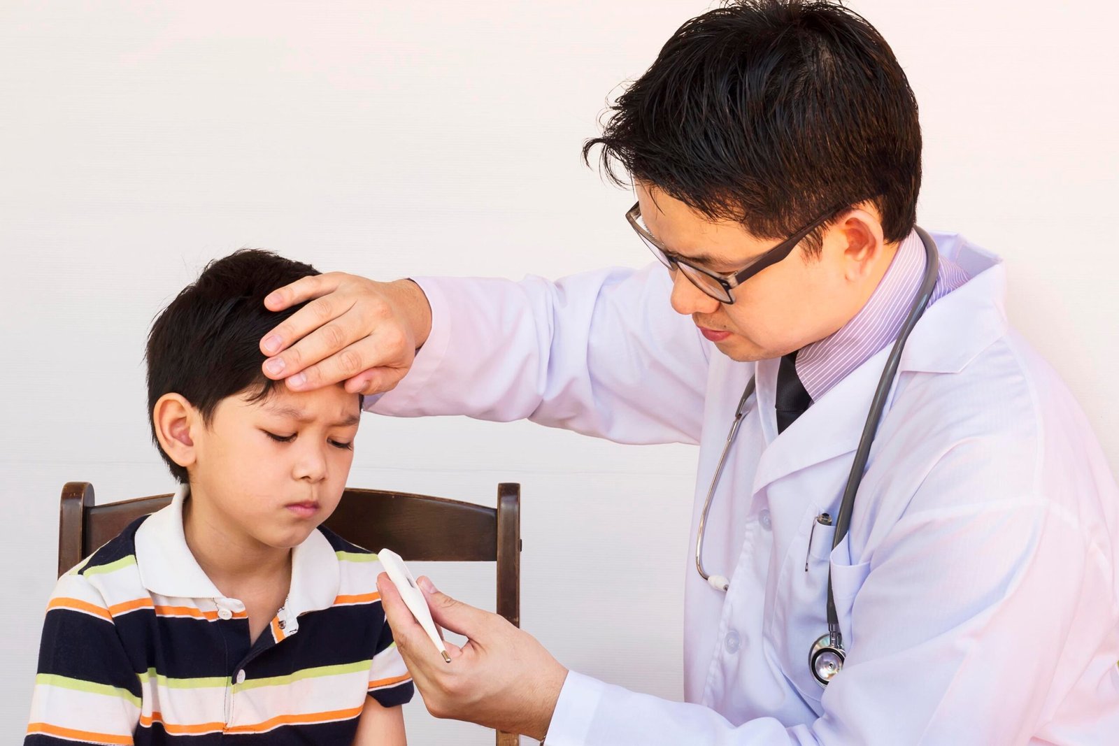
Treatment for jaundice is directed at the underlying cause. The specific treatment approach depends on whether the jaundice is pre-hepatic, hepatic, or post-hepatic in origin and whether the patient is a newborn baby or an adult.
Treatment for Jaundice in Adults
Viral Hepatitis: Hepatitis A and E are self-limiting and are managed with supportive care rest, adequate hydration, and avoidance of hepatotoxic substances including alcohol. Chronic hepatitis B and C require antiviral therapy prescribed by hepatologists at SRM Global Hospitals. Timely treatment of hepatitis B and C effectively suppresses viral replication, reduces liver injury, and prevents progression to cirrhosis.
Alcoholic Liver Disease: Complete alcohol abstinence is the cornerstone of treatment. Nutritional support, corticosteroids in severe alcoholic hepatitis, and management of complications of liver disease are provided under specialist hepatology care.
Obstructive Jaundice from Gallstones: Gallstones obstructing the bile ducts require ERCP to remove the stones endoscopically, followed by laparoscopic cholecystectomy (gallbladder removal) to prevent recurrence. Jaundice treated promptly by clearing the bile duct obstruction resolves rapidly once bile flow is restored.
Biliary Atresia: Biliary atresia in newborn babies requires early surgical intervention the Kasai portoenterostomy procedure to establish bile flow from the liver to the intestine. Early diagnosis and surgery before 60 days of life significantly improves outcomes. Liver transplantation is required for infants in whom the Kasai procedure does not achieve adequate bile flow.
Haemolytic Anaemia: Treating the underlying cause of haemolysis whether immunosuppression for autoimmune haemolytic anaemia, disease-modifying therapy for sickle cell disease, or treatment of infection in sepsis-related haemolytic disease reduces bilirubin production and resolves jaundice.
Drug-Induced Liver Injury: Stopping the offending medication under medical supervision allows the liver to recover in most cases of drug-induced liver injury. Hepatologists at SRM Global Hospitals review all medications in jaundiced patients to identify and eliminate potential hepatotoxic agents.
Treatment for Jaundice in Newborns
Phototherapy (Light Therapy)
Phototherapy is the first-line treatment for managing pathological unconjugated hyperbilirubinemia in newborns. Phototherapy works by converting bilirubin into water-soluble isomers that can be excreted through the mechanisms of photoisomerization and structural isomerization without requiring the baby’s liver to conjugate the bilirubin first. These water-soluble bilirubin isomers are excreted through the baby’s urine and bowel movements, effectively reducing total serum bilirubin levels and the risk of bilirubin toxicity.
The threshold for initiating phototherapy is determined by the infant’s gestational age, risk factors for neurotoxicity, and specific total serum bilirubin (TSB) levels with lower thresholds for preterm infants and infants with neurotoxicity risk factors.
Phototherapy lights are placed above the newborn baby in the newborn nursery. The baby’s eyes are protected with phototherapy units eye covers, and the infant’s skin is maximally exposed to the phototherapy lights to optimise bilirubin clearance. Intensive phototherapy using multiple phototherapy lights or specialised high-intensity phototherapy units is employed when total serum bilirubin levels are very high or rising rapidly.
Exchange Transfusion
Exchange transfusion is a second-line treatment for severe unconjugated hyperbilirubinemia when phototherapy is ineffective or when bilirubin levels reach critical thresholds that risk bilirubin encephalopathy. During exchange transfusion, the baby’s blood is gradually removed and replaced with donor blood physically removing bilirubin from the baby’s bloodstream and replacing the antibody-coated red blood cells in cases of blood group incompatibilities.
Intravenous Immunoglobulin (IVIG)
Intravenous immunoglobulin (IVIG) can be used in cases of immune-mediated haemolysis such as Rh or ABO incompatibility to reduce the need for exchange transfusion in newborn babies with rising bilirubin levels despite intensive phototherapy. IVIG works by blocking the immune destruction of the baby’s red blood cells, reducing the rate of haemolysis and bilirubin production.
Breastfeeding Support
For breastfeeding jaundice, supporting the mother to establish effective breastfeeding and ensuring adequate breast milk intake is the primary intervention. Frequent feeding eight to twelve times per day stimulates bowel movements and increases bilirubin excretion through the gut. Supplementation with expressed breast milk or formula may be recommended when the baby’s bilirubin level remains elevated despite feeding optimisation.
Discharge Criteria for Jaundiced Newborns
Infants treated for neonatal jaundice can be discharged when they are feeding adequately and have had two successive serum bilirubin levels demonstrating a trend toward lower values. Parents should be provided with verbal and written information about jaundice before discharge including procedures for contacting the birth hospital if the baby’s skin or eye yellow color appears to worsen after discharge.
How to Prevent Jaundice
Preventing Jaundice in Adults
While not all causes of jaundice can be prevented, several important preventive measures significantly reduce the risk:
Hepatitis B vaccination — the hepatitis B vaccine provides highly effective protection against hepatitis B infection, one of the leading causes of chronic liver disease and jaundice worldwide
Hepatitis A vaccination — recommended for travellers to endemic areas and for individuals with liver disease
Avoid excessive alcohol consumption — chronic alcohol use is a leading cause of liver disease and jaundice; limiting or eliminating alcohol protects liver function long-term
Safe food and water practices — preventing hepatitis A and E through safe food and water practices, particularly when travelling
Medication caution — avoid self-medicating with herbal preparations or medications not prescribed by a healthcare professional, as drug-induced liver injury is an important preventable cause of jaundice
Regular health screening — blood tests including liver function tests and serum bilirubin are part of routine health checks, enabling early detection of liver disease before jaundice develops
Preventing Jaundice in Newborns
Prevent jaundice complications in newborns through:
Antenatal blood group screening — identifying Rh and ABO blood group incompatibilities in pregnancy allows anti-D prophylaxis to prevent Rh haemolytic disease
Early and frequent breastfeeding — adequate feeding prevents breastfeeding jaundice by promoting bowel movements and bilirubin excretion
Universal newborn jaundice screening — assessing all newborn babies for jaundice every 12 hours as recommended, with transcutaneous bilirubin measurement or total serum bilirubin testing when jaundice is suspected
Prompt treatment — early initiation of phototherapy when the baby’s bilirubin level crosses treatment thresholds prevents progression to severe jaundice and bilirubin encephalopathy
Complications of Untreated Jaundice
The complications of jaundice depend on the underlying cause and how rapidly treatment is initiated.
In Adults
Liver failure — progressive liver disease causing jaundice can advance to acute or chronic liver failure, which is life-threatening
Portal hypertension and variceal bleeding — cirrhosis causes high pressure in the portal vein, leading to oesophageal varices that can bleed catastrophically
Hepatic encephalopathy — accumulation of toxins from liver failure affects brain function, causing confusion, disorientation, and coma
Cholangitis — untreated bile duct obstruction leads to ascending infection of the bile ducts, causing septic shock and organ failure
In Newborns
Bilirubin encephalopathy (Kernicterus) the most serious complication of untreated neonatal jaundice. Excess unconjugated bilirubin crosses the blood brain barrier and deposits in the brain, causing chronic bilirubin encephalopathy a form of permanent brain damage characterized by neurologic complications including cerebral palsy, hearing loss, intellectual disability, and movement disorders.
Kernicterus the end-stage of severe, untreated neonatal hyperbilirubinemia. Severe jaundice can lead to serious complications including brain damage if left untreated, making it critical for healthcare providers to monitor bilirubin levels closely.
Get Expert Care for Jaundice at SRM Global Hospitals — Book an Appointment
SRM Global Hospitals in Chennai provides comprehensive jaundice evaluation and management from neonatal jaundice monitoring and phototherapy in the newborn nursery to advanced hepatology and interventional gastroenterology services for adult jaundice.
Neonatal jaundice assessment and phototherapy — transcutaneous bilirubin and total serum bilirubin monitoring, phototherapy lights, and intensive phototherapy for jaundiced newborns in a fully equipped newborn nursery
Exchange transfusion services for severe neonatal hyperbilirubinemia — performed by experienced neonatologists with full intensive care support
Hepatology and gastroenterology for adult jaundice — viral hepatitis, liver disease, alcoholic liver disease, and autoimmune liver conditions
ERCP and interventional bile duct management — endoscopic removal of gallstones and stenting for obstructive jaundice
Advanced liver imaging — ultrasound, MRCP, CT, and liver biopsy for accurate diagnosis of the underlying cause
Paediatric surgery for biliary atresia — early Kasai portoenterostomy for newborn babies with bile duct obstruction
Comprehensive blood tests and liver function tests — same-day results with specialist interpretation
Multidisciplinary jaundice care — hepatologists, neonatologists, gastroenterologists, interventional endoscopists, and paediatric surgeons collaborating for every complex jaundice case
FAQs on Jaundice
1. What Are the Main Jaundice Symptoms?
The main jaundice symptom is the yellow color of the skin, whites of the eyes, and mucous membranes caused by excess bilirubin accumulating in body tissues. In adults, jaundice is typically accompanied by dark urine, pale or clay-coloured stools, itching, fatigue, and nausea. Fever and chills with jaundice suggest an active infection such as viral hepatitis or bile duct infection. In newborn babies, jaundice symptoms include yellowing of the infant’s skin starting on the face and spreading downward, poor feeding, and lethargy.
2. What Causes Jaundice?
Common conditions that cause jaundice include liver disease, viral hepatitis, gallstones blocking the bile ducts (obstructive jaundice), haemolytic anaemia causing excess bilirubin from red blood cell destruction, biliary atresia in newborn babies, and drug-induced liver injury. Jaundice can be classified into pre-hepatic (haemolytic), hepatic (liver dysfunction), and post-hepatic (bile duct obstruction) causes each requiring different treatment. Identifying the underlying cause of jaundice is the priority of every clinical evaluation.
3. What Is Newborn Jaundice and When Is It Serious?
Newborn jaundice neonatal jaundice is extremely common, affecting most newborn babies to some degree in the first week of life. Physiological jaundice is the most common type, accounting for approximately 75% of neonatal hyperbilirubinemia cases, and typically resolves within two to three weeks. However, jaundice that appears within the first 24 hours after birth, progresses rapidly, or is associated with blood group incompatibilities is pathological and requires urgent evaluation. Severe jaundice can cause bilirubin encephalopathy and serious neurologic complications if untreated making close monitoring and timely phototherapy treatment essential.
4. How Is Jaundice Treated in Newborns?
Phototherapy is the first-line treatment for pathological neonatal jaundice. Phototherapy lights convert unconjugated bilirubin in the baby’s skin into water-soluble isomers that are excreted through the bowel movements and urine without requiring liver processing. The threshold for initiating phototherapy is determined by the infant’s gestational age and neurotoxicity risk factors. Exchange transfusion is used for severe cases when phototherapy is insufficient. Intravenous immunoglobulin (IVIG) is used in immune-mediated haemolytic disease to reduce the need for exchange transfusion. For breastfeeding jaundice, ensuring adequate breast milk intake is the primary intervention.
5. What Is the Difference Between Physiological and Pathological Jaundice in Newborns?
Physiological jaundice is the normal, expected rise in bilirubin level that occurs in most newborn babies as the liver matures it appears after the first 24 hours of life, peaks at 48 to 96 hours, and resolves within two to three weeks without specific treatment beyond monitoring. Pathological jaundice appears within the first 24 hours after birth, rises more rapidly, and reflects an underlying cause such as blood group incompatibilities, haemolytic disease, biliary atresia, or sepsis requiring urgent investigation and treatment to prevent bilirubin toxicity.
6. What Do Dark Urine and Pale Stools Mean in Jaundice?
Dark urine and pale stools are commonly associated with jaundice and indicate obstructive jaundice a blockage of the bile ducts. Dark urine occurs because conjugated bilirubin is excreted through the kidneys rather than through bile. Pale or clay-coloured stools indicate that bile is blocked from reaching the digestive tract, removing the bile pigments that normally colour the stool brown. This combination strongly suggests obstruction of the bile ducts from gallstones, biliary atresia, pancreatic cancer, or other causes and requires urgent investigation including ultrasound and liver function tests.
7. Can Jaundice Be Prevented?
Jaundice from some causes can be effectively prevented. Hepatitis B vaccination prevents hepatitis B infection a leading cause of chronic liver disease and jaundice. Avoiding excessive alcohol consumption prevents alcoholic liver disease. Safe food and water practices prevent hepatitis A and E. In newborn babies, antenatal blood group screening and anti-D prophylaxis prevent severe Rh haemolytic disease. Universal newborn jaundice screening with transcutaneous bilirubin or total serum bilirubin measurement ensures jaundice is detected and treated before it progresses to severe jaundice or bilirubin encephalopathy.
