


Echo Test (Echocardiogram): Purpose, Procedure & Cost
An echo test short for echocardiogram is a non-invasive cardiac imaging procedure that uses high-frequency sound waves to create real-time images of the heart’s structure, chambers, valves, and pumping function. It is one of the most important diagnostic tools in cardiology, allowing cardiologists to evaluate the heart in motion and identify structural abnormalities, valve diseases, and weakened heart muscle that cannot be detected by an ECG test or physical examination alone.
Cardiologists and cardiac imaging specialists at SRM Global Hospitals in Chennai use the echo test as a primary tool to diagnose heart conditions, track existing conditions, and guide treatment plans for a wide range of cardiovascular diseases. The echo test delivers real time images of the heart at work providing valuable insights into cardiac structure and function within a single, painless test.
What Is an Echo Test?
An echo test also called an echocardiogram or cardiac ultrasound is a diagnostic test that uses high-frequency sound waves to produce real time images of the heart.
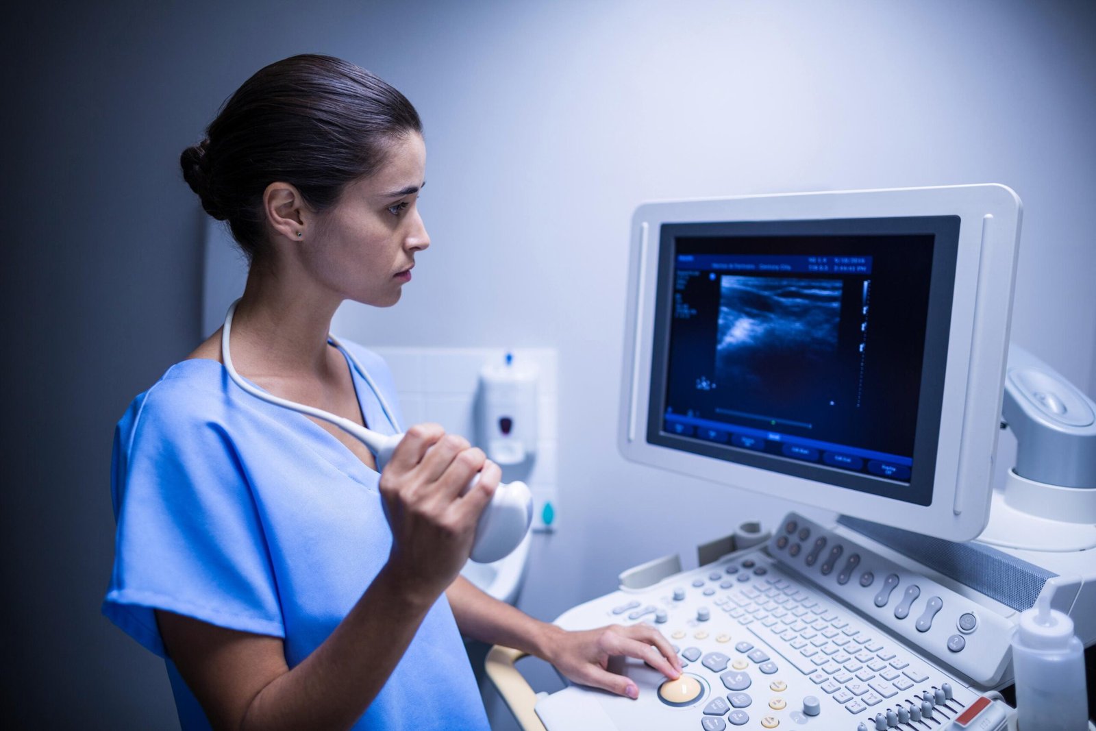
A device called a transducer sends out high-frequency sound waves (ultrasound waves) that travel into the chest area and bounce off the heart’s walls, valves, and heart tissue. These echoes are converted into live video images displayed on a computer screen, showing the heart in real time as it contracts and relaxes.
Unlike an ECG test, which records the heart’s electrical activity, the echo test creates a visual picture of the heart’s physical structure showing how the heart muscle is pumping blood, how the valves open and close, and how blood flow moves through each chamber.
The echo test is completely safe, non-invasive, and involves no radiation exposure a significant safety advantage over imaging techniques like CT scans or X-rays. The 2D echo test is considered extremely safe, with no known risks associated with the procedure, making it suitable for individuals of all ages including elderly patients and children.
An echo test is also referred to as:
Echocardiogram
Cardiac ultrasound
2D echo test (the most commonly performed type)
2D echo
Doppler echo (when blood flow assessment is included)
Transthoracic echocardiogram (TTE)
What Is a 2D Echo Test?
A 2D echo test short for two-dimensional echocardiogram is the standard form of echo test typically performed in clinical practice.
It uses ultrasound waves to produce two-dimensional cross-sectional real time images of the heart, allowing cardiologists to visualise the heart’s four chambers, the thickness and movement of the heart wall, the opening and closing of all four heart valves, and the overall efficiency of the heart muscle pumping blood.
The 2D echo is the most commonly ordered echo test at SRM Global Hospitals because it provides the clearest structural overview of the heart in the shortest examination time.
The 2D echo test does not involve radiation exposure, which is a significant advantage compared to other imaging techniques. It forms the foundation of cardiac diagnostics and is almost always combined with Doppler imaging to assess blood flow simultaneously.
The 2D echo test price in India typically ranges from ₹800 to ₹4,500, depending on the facility type, city, and whether additional Doppler and colour flow assessment are included. At SRM Global Hospitals, the 2D echo test is performed by experienced cardiologists and cardiac sonographers with same-day result reporting ensuring patients receive clinically actionable findings without delay.
Why Is an Echo Test Done?
Doctors typically request an echocardiogram to investigate concerning symptoms like unexplained chest pain and heart palpitations, as well as to evaluate early signs of heart conditions that may not be apparent from an ECG machine reading or clinical examination alone.
Cardiologists at SRM Global Hospitals recommend an echo test when a patient’s symptoms or ECG results suggest the possibility of structural heart disease or impaired cardiac function. The echo test provides valuable insights about the heart’s structure and mechanical performance that no other single cardiac diagnostic test can deliver.
Common Reasons for an Echo Test
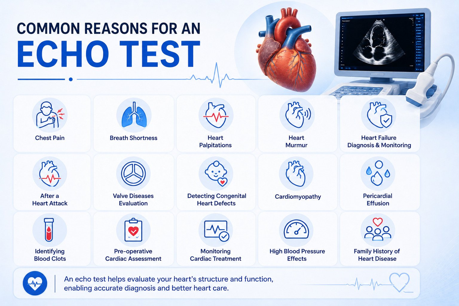
Chest pain — to evaluate whether valve diseases, pericardial effusion, or wall motion abnormalities are contributing to symptoms, and to directly detect angina or coronary artery disease
Breath shortness — to assess whether weakened heart muscle or heart failure is the underlying cause of breathing difficulty
Heart palpitations — to identify whether structural abnormalities or valve disorders are causing irregular heartbeats
Heart murmur — to identify the origin, severity, and haemodynamic significance of an abnormal heart sound, and to assess valve function precisely
Heart failure diagnosis and monitoring — to measure ejection fraction and assess how effectively the heart muscle is pumping blood, tracking disease progression over time
After a heart attack — to assess the extent of heart tissue damage, identify weakened heart muscle, and detect any complications such as fluid accumulation around the heart
Valve diseases evaluation — to assess the severity of mitral stenosis, mitral regurgitation, aortic stenosis, aortic regurgitation, and other valve disorders that affect blood flow
Detecting congenital heart defects and birth defects — to identify structural abnormalities and congenital defects present from birth, including septal defects and abnormal valve formation
Cardiomyopathy — to evaluate abnormal thickening, heart enlargement, or weakened heart muscle
Pericardial effusion — to detect fluid accumulation and fluid buildup around the heart
Identifying blood clots — the echo test can identify blood clots within heart chambers that carry a risk of stroke
Pre-operative cardiac assessment — to establish a baseline of cardiac structure and function before heart surgery or major surgical procedures
Monitoring cardiac treatment — to assess whether medications or interventions have improved heart structure and function, providing valuable insights into treatment response
High blood pressure effects — to detect left ventricular hypertrophy or diastolic dysfunction caused by long-standing blood pressure elevation, particularly in elderly patients
Family history of heart disease — to screen for early signs of inherited cardiac abnormalities in patients with a significant family history of heart conditions
When Should You Get an Echo Test Done?
Symptom or Clinical Finding | Why an Echo Test Is Needed |
|---|---|
Unexplained breath shortness | To assess heart muscle pumping function and rule out heart failure |
Chest pain with normal ECG machine reading | To detect wall motion abnormalities or pericardial effusion |
Heart murmur on examination | To identify and grade valve diseases and valve disorders |
After a heart attack | To assess heart tissue damage and ejection fraction |
Persistent high blood pressure | To detect heart enlargement and diastolic dysfunction |
Heart palpitations with dizziness | To identify structural abnormalities or congenital defects |
Suspected blood clots in heart chambers | To capture images of intracardiac thrombus |
Family history of heart conditions | To detect early signs of inherited structural abnormalities |
Types of Echo Tests
Cardiologists and cardiac imaging specialists at SRM Global Hospitals select the most appropriate type of echo test based on the clinical question, the patient’s anatomy, and the image clarity required.
Transthoracic Echocardiogram (TTE) — 2D Echo Test
The transthoracic echocardiogram commonly called the 2D echo test is the standard, most widely and typically performed echo test.
A device called a transducer is placed on the chest wall over specific positions on the chest area, and high-frequency sound waves are directed through the chest to create real time images of the heart on a computer screen.
The 2D echo test simultaneously captures:
Two-dimensional (2D) imaging — structural real time images of heart chambers, walls, and valves
M-mode imaging — precise measurements of chamber dimensions and wall thickness, within the normal range for the patient’s age and body size
Doppler imaging — assessment of blood flow velocity and direction through heart valves and chambers
Colour Doppler — colour-coded visualisation of blood flow patterns, identifying valve disorders such as regurgitation or stenosis
The TTE is non-invasive, requires no sedation, and is typically performed and completed within 15 to 30 minutes at SRM Global Hospitals.
Transoesophageal Echocardiogram (TOE/TEE)
Transoesophageal echocardiography (TEE) involves inserting a probe into the oesophagus to obtain detailed heart images, particularly useful for visualising posterior heart structures with greater image clarity than a standard 2D echo.
The TEE is used when the standard 2D echo test does not provide sufficient detail, when evaluating the mitral valve before heart surgery, when assessing blood clots in the left atrial appendage, or when investigating infective endocarditis. While the chances of complications from a 2D echo test are almost nil, rare complications can occur with TEE including mild throat irritation or nausea during the procedure. Cardiac imaging specialists at SRM Global Hospitals perform TOE under mild sedation in a controlled clinical setting.
Stress Echocardiogram
Stress echocardiography is performed while the heart is under physical stress either through exercise on a treadmill test or exercise bike, or through pharmacological stress using medications that simulate the effects of exercise. This allows cardiologists to assess how the heart responds under increased workload and is useful for identifying coronary artery disease, wall motion abnormalities, and valve disorders that only become apparent when cardiac demand is elevated.
The stress echocardiogram is particularly useful for patients who cannot undergo nuclear stress testing and for those where the healthcare provider needs to directly detect angina or assess functional significance of valve diseases.
Doppler Echocardiography?
Doppler echocardiography specifically examines blood flow through the heart and is instrumental in diagnosing cardiac abnormalities such as valve leaks or blockages. Doppler ultrasound is used to track the velocity and direction of blood flow through the heart valves providing valuable insights into the pressure gradients across stenotic valves, the severity of regurgitation, and any abnormal blood flow patterns caused by congenital defects or structural abnormalities.
Three-Dimensional (3D) Echocardiogram
A 3D echocardiogram uses advanced ultrasound technology to capture images in three dimensions, providing more accurate measurements of heart chamber volumes and ejection fraction than standard 2D echo imaging.
It is particularly valuable for complex valve repair planning before heart surgery and for structural heart interventions where precise volumetric data is essential. Cardiac imaging specialists at SRM Global Hospitals use 3D echocardiography for selected cases requiring detailed surgical or interventional planning.
Contrast Echocardiogram
A contrast echocardiogram involves injecting a small amount of contrast dye (microbubble contrast agent) intravenously to improve image clarity of left ventricular borders when standard 2D echo images are suboptimal.
It provides more accurate ejection fraction measurements and helps identify apical wall motion abnormalities or intracardiac masses within the heart chambers. The contrast dye used is different from that used in CT scans and carries minimal risk.
How Is a 2D Echo Test Performed?
The echo test procedure at SRM Global Hospitals is straightforward, painless, and typically completed within 15 to 30 minutes. No special preparation is required for a standard transthoracic 2D echo test.
Before the Echo Test
No fasting is required for a standard resting 2D echo test.
Patients may eat, drink, and take regular medications normally before the appointment. For a stress echocardiogram, patients are advised to avoid heavy meals for two to three hours before and wear comfortable clothing for the treadmill test.
For a transoesophageal echocardiogram, fasting for four to six hours is required, and a responsible adult must accompany the patient for discharge after sedation. Patients should inform the healthcare provider about their complete medical history, including previous heart surgery, pacemaker implantation, or known valve diseases.
During the 2D Echo Test — Step by Step
Positioning: The patient lies on an examination table, typically on their left side, which brings the heart closer to the chest wall and improves image clarity.
ECG electrode placement: Electrodes are placed on the chest to track the heart’s electrical activity and rhythm on the ECG machine simultaneously during the echo scan.
Gel application: A clear, water-based ultrasound gel is applied to the chest area to facilitate sound wave transmission and eliminate air pockets that would block the high-frequency sound waves.
Transducer placement: The cardiac sonographer places a device called a transducer at specific positions on the chest area parasternal, apical, subcostal, and suprasternal windows to capture images from multiple angles.
Image capture: Real time images, Doppler measurements, and colour flow maps are captured and recorded. The transducer sends out high-frequency sound waves that bounce off the heart’s walls and valves, converting these echoes into live video on a computer screen.
Reporting: The cardiologist reviews the heart images and measurements and generates a comprehensive echo test report, typically available the same day at SRM Global Hospitals.
What to Expect During the Echo Test
The procedure is a painless test from start to finish.
Patients feel only a slight pressure of the transducer on the chest area and the slight coolness of the ultrasound gel no pain, no discomfort. The ultrasound gel facilitates sound wave transmission and is wiped off completely at the end of the procedure.
No radiation, no needles, and no contrast dye are required for a standard 2D echo test. Patients remain awake and can communicate with the sonographer throughout. The echo test is typically completed within 15 to 30 minutes, after which patients can leave within a few hours of their appointment with no recovery period needed.
2D Echo Test Price
The cost of a 2D echo test in India typically ranges from ₹800 to ₹4,500, depending on various factors such as the type of facility, the specific location within India, and any additional services required during the test.
In some locations, the 2D echo test price can be as low as ₹850, while at private multi-specialty hospitals, it can go up to ₹2,000 or more depending on whether Doppler imaging, colour flow mapping, and cardiologist interpretation are included. Factors affecting the price of a 2D echo test include the type of facility, location, and additional services required.
Patients at SRM Global Hospitals are advised to contact the cardiac diagnostics team directly for current 2D echo test pricing, health package options, and health insurance coverage details.
Understanding Echo Test Results
Echo test results are reported by experienced cardiologists at SRM Global Hospitals as a structured report covering all key parameters of cardiac structure and function. Understanding the main measurements helps patients engage meaningfully with their diagnosis and treatment plan.
Key Echo Test Measurements and Normal Range
Measurement | What It Evaluates | Normal Range (Adult) |
|---|---|---|
Ejection Fraction (EF) | Percentage of blood pumped out of the left ventricle with each beat reflects pumping blood efficiency | 55% – 70% |
Left Ventricular Internal Diameter (LVID) | Size of the left ventricle chamber | Diastole: 3.9–5.3 cm |
Interventricular Septal Thickness (IVS) | Thickness of the wall between ventricles elevated in hypertrophy | 0.6–1.1 cm |
Left Atrial Size | Diameter of the left atrium enlarged in atrial fibrillation or mitral valve diseases | Less than 4.0 cm |
Aortic Root Diameter | Width of the aorta at its root | 2.0–3.7 cm |
E/A Ratio (Mitral Doppler) | Diastolic function assesses how well the heart relaxes and fills with blood | Greater than 1.0 in normal adults |
Peak Gradient (Valve Doppler) | Pressure difference across a valve elevated in valve stenosis | Valve-specific |
What Echo Test Results Can Detect
Echocardiograms evaluate pumping strength by measuring the ejection fraction, detect valve problems and valve disorders, identify heart enlargement, find congenital defects and birth defects, identify blood clots, assess damage after heart attacks, measure heart chamber sizes, and identify fluid accumulation around the heart. The echo test is a primary tool used to diagnose heart diseases, track existing conditions, and guide treatment plans providing a complete picture of cardiac health in a single non-invasive test.
Common Abnormal Echo Test Findings
Reduced ejection fraction (below 50%): Indicates weakened heart muscle and systolic heart failure the heart is not pumping blood efficiently. Cardiologists at SRM Global Hospitals classify this further as mildly, moderately, or severely reduced to guide treatment intensity.
Left ventricular hypertrophy (LVH): Heart enlargement and abnormal thickening of the left ventricular wall commonly caused by long-standing blood pressure elevation or hypertrophic cardiomyopathy.
Wall motion abnormality: Segments of the heart wall that move abnormally indicating poor blood flow from coronary artery disease or previous heart attack damage to heart tissue.
Valve diseases — mitral stenosis: Restricted opening of the mitral valve, causing elevated pressures in the left atrium. Severity is graded by valve area and pressure gradient on Doppler.
Valve diseases — mitral regurgitation: Backward leaking of blood through the mitral valve, graded mild, moderate, or severe by colour Doppler.
Valve diseases — aortic stenosis: Calcified, narrowed aortic valve with high Doppler gradients one of the most common valve disorders in elderly patients, potentially requiring valve replacement via heart surgery.
Pericardial effusion: Fluid accumulation and fluid buildup around the heart. A large pericardial effusion can compress the heart and requires urgent intervention.
Detecting congenital heart defects: The 2D echo is the primary tool for detecting congenital heart defects and birth defects including septal defects, valve abnormalities, and other congenital defects present from birth.
Dilated cardiomyopathy: Heart enlargement with globally weakened heart muscle and reduced ejection fraction.
Hypertrophic cardiomyopathy (HCM): Abnormal thickening of the interventricular septum, detected on 2D echo as asymmetric hypertrophy a cause of sudden cardiac events in younger patients and athletes.
Intracardiac blood clots: Blood clots within heart chambers, identified as echogenic masses carrying significant stroke risk.
Abnormal Findings and Further Testing
An abnormal echo test provides the healthcare provider with precise structural information to guide the next step in management. Abnormal findings often require further testing to confirm severity and plan treatment.
Depending on the findings, experienced cardiologists at SRM Global Hospitals may recommend:
Optimisation of heart medications such as ACE inhibitors, beta blockers, or diuretics for weakened heart muscle or heart failure
Serial echo tests to monitor progression of valve diseases or cardiomyopathy over time
Referral to interventional cardiology for valve repair or replacement before further heart surgery is required
Coronary angiography if wall motion abnormalities and poor blood flow suggest significant coronary artery disease
Cardiac surgery consultation for complex structural abnormalities
Urgent drainage if pericardial effusion with haemodynamic compromise is identified
Echo Test vs. ECG Test: Key Differences
Many patients ask how an echo test differs from an ECG test. Both are essential cardiac diagnostic tools but they measure completely different aspects of heart function.
Feature | Echo Test (Echocardiogram) | ECG Test (Electrocardiogram) |
|---|---|---|
What it measures | Heart structure, chamber size, valve function, ejection fraction | Heart’s electrical activity, rhythm, conduction |
Technology used | High-frequency sound waves (ultrasound) | Electrodes recording electrical activity |
What it detects | Valve diseases, heart failure, congenital defects, wall motion abnormality | Arrhythmias, heart attack, conduction defects |
Duration | 15–30 minutes | 5–10 minutes |
Radiation | None | None |
Computer screen output | Real time images of heart structure | Waveform tracing of electrical activity |
When ordered together | When structural and electrical assessment are both needed | When rhythm and electrical evaluation is the priority |
Cardiologists at SRM Global Hospitals frequently order both an echo test and an ECG test together using the ECG machine reading alongside 2D echo real time images to obtain a complete picture of cardiac structure, electrical activity, and overall heart health.
Get a Complete Cardiac Assessment — Book an Appointment
SRM Global Hospitals in Chennai provides comprehensive echo testing and cardiac diagnostics, including 2D echo test, stress echocardiogram, transoesophageal echocardiogram, Doppler echocardiography, and 3D echocardiography all reported by experienced cardiologists with same-day results.
Standard 2D echo test with Doppler and colour flow mapping same-day reporting by experienced cardiologists for accurate real time images and results
Stress echocardiogram using treadmill test or pharmacological stress for detection of coronary artery disease and wall motion abnormalities
Transoesophageal echocardiogram (TOE) for high-resolution image clarity of posterior cardiac structures and valve diseases assessment before heart surgery
Doppler echocardiography to track velocity and direction of blood flow through heart valves and detect valve disorders
3D echocardiography for complex valve repair planning and precise volumetric measurements of heart chambers
Contrast echocardiogram with contrast dye for accurate ejection fraction assessment when standard 2D echo image clarity is suboptimal
Integrated cardiac diagnostics — echo test findings correlated with ECG machine results, coronary angiography, and cardiac biomarkers
Transparent 2D echo test pricing with health package options, insurance-friendly billing, and clear 2D echo test price information
FAQs on Echo Test
1. What Is an Echo Test and What Does It Show?
An echo test or echocardiogram is a non-invasive diagnostic test that uses high-frequency sound waves to create real time images of the heart’s structure on a computer screen. A device called a transducer sends out ultrasound waves that bounce off the heart tissue and heart chambers, converting the echoes into live video graphics showing heart valves, pumping blood function, and blood flow in real time. The echo test is a primary tool used to diagnose heart conditions, track existing conditions, and guide treatment plans providing valuable insights into the heart’s structural health.
2. What Is a 2D Echo Test?
A 2D echo test or two-dimensional echocardiogram is the standard form of echo test typically performed in clinical practice. It uses high-frequency sound waves to produce two-dimensional real time images of the heart’s structure and function. The 2D echo test captures real time images of all four heart chambers, the heart valves, and surrounding heart tissue combined with Doppler imaging to assess blood flow simultaneously. It is the most commonly performed cardiac imaging test at SRM Global Hospitals and is considered extremely safe with no known risks for patients of all ages.
3. What Is the 2D Echo Test Price in India?
The cost of a 2D echo test in India typically ranges from ₹800 to ₹4,500 depending on the facility type, city, and services included. In some locations, the 2D echo test price can be as low as ₹850, while at private multi-specialty hospitals it can go up to ₹2,000 or more when Doppler imaging and cardiologist interpretation are included. Factors affecting the price of a 2D echo test include location, facility type, and additional services required. Patients at SRM Global Hospitals are advised to contact the cardiac diagnostics team directly for current pricing and health package options.
4. Is an Echo Test Painful or Harmful?
The echo test is a painless test with no known risks for a standard 2D echo. No radiation exposure is involved a significant advantage over CT scans or X-rays. Patients feel only a slight pressure from the transducer on the chest area and the coolness of the ultrasound gel applied for sound wave transmission. No needles, no contrast dye, and no sedation are required for a transthoracic 2D echo. Rare complications can occur with transoesophageal echocardiogram (TEE) mild throat irritation or nausea but the standard 2D echo test carries no such risks.
5. How Long Does an Echo Test Take?
A standard 2D echo test typically lasts between 15 to 30 minutes, including image capture, Doppler measurement, and initial reporting. Patients can leave within a few hours of the appointment with no recovery time needed. A stress echocardiogram, which includes a treadmill test, takes approximately 60 to 90 minutes. Same-day echo test reports are available at SRM Global Hospitals for most echo test types.
6. What Heart Conditions Can an Echo Test Detect?
Echocardiograms can detect a wide range of heart conditions and cardiac abnormalities. These include valve diseases and valve disorders, heart failure and weakened heart muscle, detecting congenital heart defects and birth defects, identifying blood clots in heart chambers, pericardial effusion and fluid accumulation, heart enlargement and cardiomyopathy, wall motion abnormalities from heart attacks, and coronary artery disease through a stress echocardiogram. The echo test is a primary tool used to diagnose heart diseases at early signs, track existing conditions, and guide treatment plans.
7. What Does a Normal Echo Test Result Mean?
A normal echo test result means the heart chambers are within the normal range for size, the heart muscle is contracting with normal strength (ejection fraction above 55%), all four valves are functioning without significant valve diseases, there is no fluid accumulation around the heart, and no structural abnormalities are detected. A normal 2D echo test provides strong reassurance about the heart’s mechanical function. The healthcare provider will correlate normal echo findings with the patient’s symptoms and medical history to determine whether further testing is warranted.
8. Who Needs an Echo Test?
Anyone experiencing unexplained breath shortness, chest pain, heart palpitations, or dizziness may be referred for an echo test. Patients with known heart failure, valve diseases, cardiomyopathy, or a history of heart attack require regular echo tests to track their condition. Patients with long-standing blood pressure issues benefit from an echo test to detect early signs of heart enlargement or diastolic dysfunction. Patients with a family history of heart conditions or congenital defects may also be screened. The healthcare provider or cardiologist at SRM Global Hospitals will assess whether an echo test is the appropriate next step in cardiac diagnostics.
9. What Is the Difference Between a Transthoracic and Transoesophageal Echo Test?
Transthoracic Echocardiography (TTE) the 2D echo test is typically performed by placing the transducer on the chest wall, making it a straightforward, painless, non-invasive test requiring no sedation. It is the standard first-line echo test for evaluating heart conditions. Transoesophageal Echocardiography (TEE) involves inserting a probe into the oesophagus to obtain detailed heart images of posterior heart structures such as the mitral valve and left atrium, with superior image clarity. TOE is performed under mild sedation and is used when the standard 2D echo test does not provide sufficient detail or when greater precision is required for heart surgery planning.
