


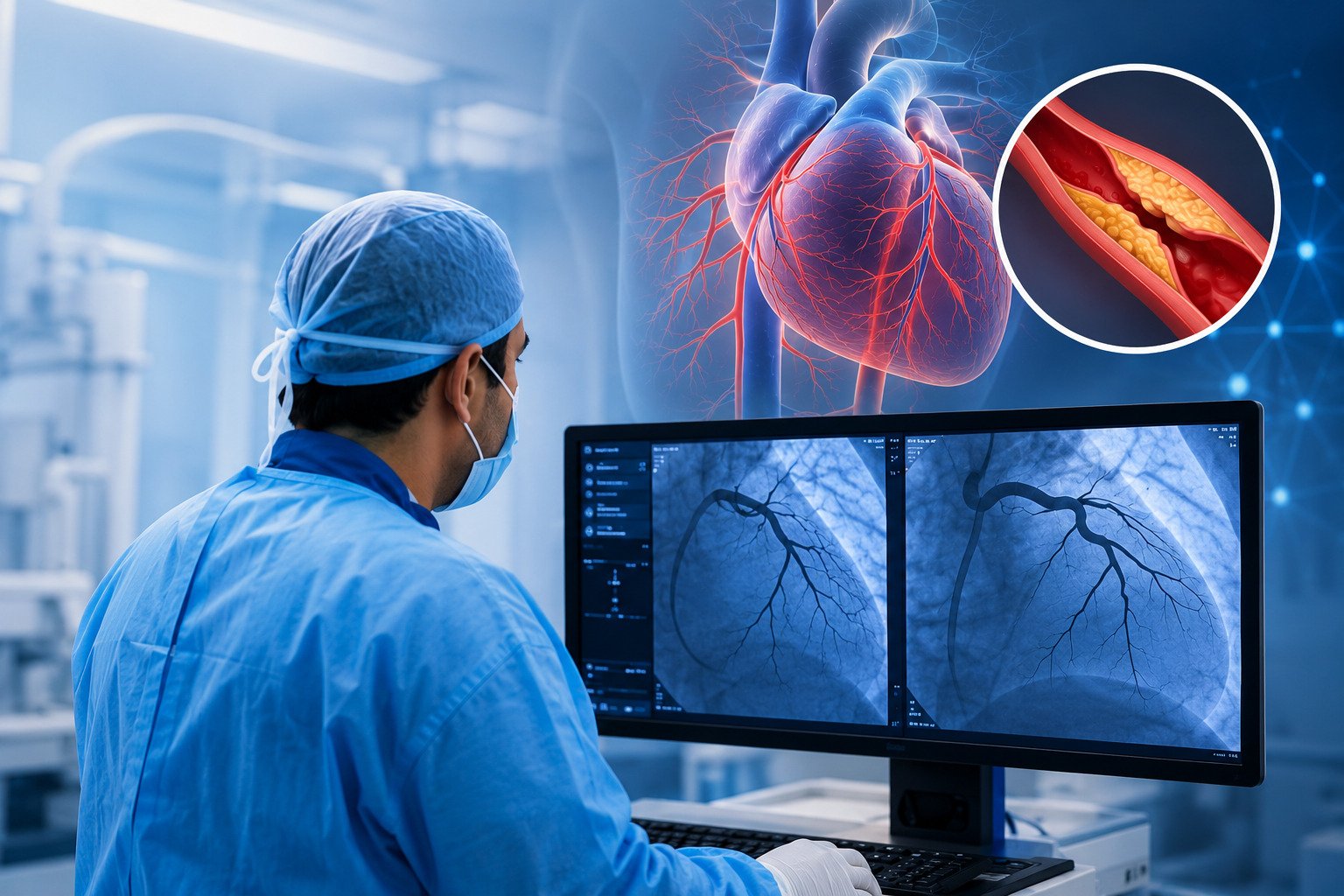
What Is an Angiogram? Procedure, Risks & Recovery
An angiogram is a diagnostic imaging procedure used to examine blood vessels, identify blockages, and assess blood flow to the heart and other vital organs. It is one of the most accurate diagnostic tools available for evaluating coronary artery disease and other cardiovascular conditions. Interventional cardiologists and cardiac imaging specialists at SRM Global Hospitals in Chennai use the coronary angiogram as a definitive diagnostic tool to map arterial anatomy, detect narrowed or blocked coronary arteries, and guide personalised treatment decisions that protect long-term heart health.
What Is an Angiogram?
An angiogram is a procedure in which a special contrast dye is injected into the blood vessels while x-ray images are captured in real time. These x-ray images allow interventional cardiologists to visualise the coronary arteries clearly and identify any narrowed artery, plaque buildup, or complete blockage that is restricting blood flow to the heart muscle.
The coronary angiogram belongs to a broader category of tests called cardiac catheterisation and invasive coronary angiography. It is the gold standard for diagnosing known coronary artery disease, assessing coronary stenosis, and determining whether blood flow to the heart is critically reduced. When performed to evaluate the coronary arteries specifically, the procedure is referred to as coronary angiography or invasive coronary angiography.
There are two primary types of angiograms used at SRM Global Hospitals:
Conventional Coronary Angiography (Invasive Procedure): A thin, flexible tube called a catheter also referred to as a thin tube called a catheter is inserted through the wrist or groin artery and guided to the heart. Contrast dye is injected directly into the coronary arteries, and x-ray images are captured to create detailed images of the arterial structure.
CT Coronary Angiogram (CTCA): A non-invasive form of computed tomography angiography that uses a CT scanner and contrast dye injected through an IV to create detailed images of the coronary arteries without catheter insertion. The CT coronary angiogram is particularly effective at identifying plaque buildup along the artery walls and assessing heart disease risk.
Both approaches give cardiologists at SRM Global Hospitals the clinical information needed to determine whether medical management, lifestyle changes, angioplasty, or bypass surgery is required.
Why Is an Angiogram Test Ordered?
Cardiologists at SRM Global Hospitals recommend an angiogram test when symptoms or non-invasive cardiac tests suggest the possibility of reduced blood flow to the heart or brain. The procedure is considered safe and effective and is recommended only when the benefits outweigh the procedural risks for each individual patient.
Common clinical reasons for a coronary angiogram include:
Acute chest pain or stable chest pain that has not been adequately explained by a stress test or other non-invasive assessment
Difficulty breathing on exertion that may indicate compromised blood flow to the heart muscle
Abnormal stress test results suggesting reduced blood supply to the coronary arteries
After a myocardial infarction (heart attack) to assess the condition of the coronary arteries and identify the site of blockage
Known coronary artery disease requiring detailed mapping before surgical or interventional planning
High blood pressure with cardiac symptoms where coronary involvement needs to be ruled out
High cholesterol with cardiac risk factors where coronary stenosis needs direct assessment
Planned cardiac surgery such as bypass grafting, where preoperative coronary angiography guides the surgical team
Unexplained irregular heartbeats associated with suspected structural or vascular heart disease
Symptoms That May Lead to an Angiogram Referral
Symptom | Why an Angiogram May Be Needed |
Persistent chest pain or acute chest pain | To rule out significant coronary artery blockage causing reduced blood flow |
Difficulty breathing on mild exertion | May indicate compromised cardiac output from a narrowed artery |
Palpitations with fainting | To evaluate structural or vascular causes affecting heart muscle function |
Arm or jaw pain radiating from the chest | Classic pattern of ischaemic heart disease requiring coronary angiography confirmation |
Abnormal ECG or echo findings | To correlate cardiac imaging findings with coronary artery anatomy |
Elevated high cholesterol with cardiac symptoms | To assess whether plaque buildup has caused coronary stenosis |
Types of Angiogram
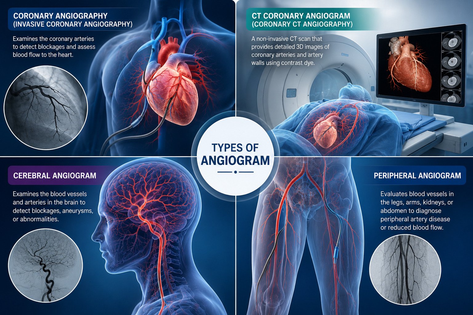
Different types of angiograms are used depending on the area of the body and the clinical question being investigated. Cardiologists, neurologists, and vascular surgeons at SRM Global Hospitals select the most appropriate type based on the patient’s medical history, symptoms, and diagnostic accuracy requirements.
Coronary Angiography (Invasive Coronary Angiography)
Coronary angiography also called invasive coronary angiography is the definitive diagnostic tool for coronary artery disease. It directly visualises the coronary arteries supplying the heart muscle and identifies any narrowed artery, coronary stenosis, or complete blockage that is restricting blood flow. The diagnostic accuracy of coronary angiography is unmatched for identifying the precise location and severity of blockages.
The results from coronary angiography directly inform whether a patient requires angioplasty with stent placement, coronary artery bypass grafting (CABG), or medical therapy with lifestyle changes. When coronary angiography shows a significant blockage, interventional cardiologists at SRM Global Hospitals can proceed directly to treatment such as angioplasty in the same session, improving blood flow to the heart immediately.
CT Coronary Angiogram (Coronary CT Angiography)
A CT coronary angiogram also called coronary CT angiography or coronary computed tomography angiography is a non-invasive diagnostic test that uses a CT scanner combined with contrast dye injected through an IV to create detailed three-dimensional images of the coronary arteries and artery walls.
The CT coronary angiogram offers unique capabilities compared to invasive coronary angiography:
Plaque characterisation: CT coronary angiography can distinguish between calcified and non-calcified plaque buildup within the artery walls, which is important for assessing heart attack risk even before a narrowed artery develops
Non-invasive assessment: No catheter site, no catheter insertion, and no arterial puncture required
Shorter procedure time: Typically completed within 15 to 30 minutes
High diagnostic accuracy for ruling out significant coronary artery disease in low-to-intermediate risk patients
Detailed anatomical mapping of coronary artery anatomy before cardiac surgery
Before a CT coronary angiogram, patients may be given medications such as beta blockers to slow their heart rate, which improves image quality and diagnostic accuracy. Patients are also generally advised to avoid caffeine for at least 12 hours before the test, as it can increase heart rate and affect the clarity of the images obtained during the CT angiogram.
Radiologists and cardiologists at SRM Global Hospitals use advanced CT coronary angiography technology to deliver high-resolution images with minimal radiation exposure.
Cerebral Angiogram
A cerebral angiogram examines the blood vessels and arteries supplying the brain. Neurologists and neurovascular specialists at SRM Global Hospitals order this test when a patient presents with stroke symptoms, unexplained severe headaches, or suspected cerebral aneurysm.
Peripheral Angiogram
A peripheral angiogram evaluates blood vessels in the legs, arms, kidneys, or abdomen. Vascular surgeons at SRM Global Hospitals use this type of angiogram to investigate peripheral artery disease, limb ischaemia, renal artery stenosis, or reduced blood flow to the extremities.
How Is a Coronary Angiogram Performed? Step-by-Step
Understanding exactly what happens during a coronary angiography procedure helps patients feel confident and prepared. The procedure is performed in a dedicated cardiac catheterisation laboratory (cath lab) at SRM Global Hospitals.
Before the Procedure
The healthcare team at SRM Global Hospitals provides detailed preparation instructions during the pre-procedure consultation. It is important for patients to inform their healthcare provider about all medications they are taking, as some medications particularly blood thinners may need to be paused or adjusted before the angiogram.
General preparation guidelines include:
Fasting: Patients are generally advised to avoid solid food for at least six hours before a coronary angiogram. Clear liquids may be allowed up to two hours prior to the procedure, as directed by the cardiologist.
Caffeine restriction: Avoid caffeine for at least 12 hours before the test, particularly before a CT coronary angiogram, as caffeine raises heart rate and reduces image clarity.
Medication review: The healthcare provider will review the patient’s complete medical history and medication list. Blood thinners, diabetes medications, and certain supplements may require temporary adjustment hours before the test.
Beta blocker administration: Before a CT coronary angiogram, the healthcare team may administer a medicine called a beta blocker to slow the heart rate and improve the quality of cardiac imaging.
Allergy disclosure: Inform the medical team of any known allergies to contrast dye, iodine, shellfish, or any medications. Pre-medication is given when necessary to prevent an allergic reaction.
Sedation preparation: Patients may receive a sedative before the procedure to help them relax, and the insertion site is cleaned and prepared with local anaesthesia.
IV line placement: An intravenous (IV) line is inserted for fluid and medication administration throughout the procedure.
Arrange transport: Patients must not drive after the procedure. A family member or friend should accompany the patient home.
During the Procedure
The patient is positioned on an x-ray table in the cath lab wearing a hospital gown and connected to cardiac monitoring equipment.
Local anaesthesia is applied to the access site most commonly the radial artery at the wrist, or alternatively the femoral artery in the groin.
A thin, flexible tube called a catheter is inserted through the access site and carefully guided through the blood vessels to the heart under continuous x-ray guidance (fluoroscopy).
Contrast dye also called contrast agent is injected through the catheter directly into the coronary arteries.
X-ray images are captured in real time as the contrast dye flows through the coronary arteries, clearly revealing any narrowed artery, plaque buildup, reduced blood flow, or complete blockage.
Interventional cardiologists at SRM Global Hospitals review the x-ray images immediately. If coronary angiography shows a significant blockage, they may proceed directly to angioplasty and stenting in the same session to restore blood flow.
The catheter is removed, and pressure is applied to the catheter site to prevent bleeding.
The entire coronary angiography procedure typically takes 30 to 60 minutes. Patients remain awake throughout, though sedation is available for anxious patients.
After the Procedure Discharge and Immediate Recovery
After the procedure, patients are moved to a monitored recovery area. Most patients can go home within four to six hours after a coronary angiogram, but some may need to stay overnight depending on their condition and whether an intervention such as angioplasty was performed.
The healthcare team at SRM Global Hospitals monitors:
The catheter site for any bleeding, swelling, or haematoma formation
Heart rate and blood pressure for stability
Kidney function if contrast dye volume was significant
Any delayed allergic reaction to the contrast agent
CT Coronary Angiogram vs. Conventional Coronary Angiography: Key Differences
Feature | CT Coronary Angiogram | Conventional Coronary Angiography |
Type of procedure | Non-invasive | Invasive procedure (catheter required) |
Anaesthesia | Not required | Local anaesthesia at catheter site |
Procedure time | 15–30 minutes | 30–60 minutes |
Hospitalisation | Usually outpatient | 4–6 hours or overnight |
Plaque assessment | Calcified and non-calcified plaque in artery walls | Primarily lumen narrowing |
Ability to treat during test | No | Yes angioplasty and stenting in same session |
Pre-test preparation | Beta blockers, caffeine restriction 12 hours before | Fasting, blood thinners adjustment |
Best suited for | Ruling out disease in lower-to-intermediate risk patients | Definitive diagnosis and intervention |
Radiation exposure | Low-dose CT radiation | Fluoroscopy radiation |
Cardiologists at SRM Global Hospitals recommend the most appropriate approach based on the patient’s risk profile, symptoms, medical history, and prior test results.
Angiogram Risks and Complications
Coronary angiography is a well-established invasive procedure with a strong safety profile. Serious complications occur in less than 1% of cases when performed by experienced interventional cardiologists in a fully equipped cath lab. The healthcare team at SRM Global Hospitals discusses all potential risks and benefits with each patient during the informed consent process before proceeding.
Common and Minor Side Effects
Bruising or soreness at the catheter site the most common minor complication, typically resolving within a few days
Temporary warm or flushing sensation when the contrast dye is injected normal and short-lived
Mild nausea in some patients following contrast agent administration
Minor bleeding at the insertion site managed with direct pressure and prevent bleeding protocols at the catheter site
Kidney stress from contrast dye patients are advised to drink plenty of water after the angiogram to help flush the contrast dye from their system and protect kidney function
Less Common Risks
Allergic reaction to contrast dye: Mild reactions such as skin rashes can occur. Patients with known sensitivities to iodine are assessed carefully, and pre-medication is given when necessary.
Kidney damage: The contrast agent is filtered through the kidneys. Patients with pre-existing kidney disease require careful hydration and monitoring. Nephrologists at SRM Global Hospitals collaborate in such cases to protect renal function.
Radiation exposure: Both conventional coronary angiography and CT coronary angiography involve some radiation exposure. Cardiologists at SRM Global Hospitals apply clinical practice guidelines to use the lowest effective dose.
Irregular heartbeats (arrhythmia) during catheter manipulation usually brief and self-resolving.
Blood clots: Although rare, blood clot forms during catheter navigation are possible. Experienced cardiologists follow strict anticoagulation protocols to minimise this risk.
Rare but Serious Complications
More serious complications from coronary angiography, although uncommon, can include:
Heart attack (myocardial infarction) caused by arterial disruption or blood clot forms during the procedure
Stroke due to dislodgement of plaque or blood clots into the cerebral blood vessels
Damage to blood vessels at the catheter site or during catheter navigation
Significant allergic reaction (anaphylaxis) to the contrast agent managed immediately with emergency medications
The risk of complications during coronary angiography may be higher in patients with certain pre-existing conditions, such as kidney disease, severe allergies to contrast dye, or advanced cardiovascular disease. Cardiologists at SRM Global Hospitals carefully evaluate each patient’s individual risk before recommending the procedure.
Who Should Exercise Caution?
Patients with the following conditions are carefully assessed by the healthcare team at SRM Global Hospitals before proceeding with conventional coronary angiography:
Severe kidney disease or significant kidney damage risk
Uncontrolled bleeding disorders or active use of blood thinners that cannot be safely paused
Severe allergy to contrast agent or iodine
Active infection or fever at the time of the procedure
In such cases, a CT coronary angiogram or an alternative cardiac imaging strategy may be preferred to supply blood flow information with lower procedural risk.
Angiogram Results: What They Mean for Your Treatment
Once the procedure is complete, interventional cardiologists at SRM Global Hospitals review the coronary angiogram findings to assess the degree of coronary stenosis and plan the appropriate treatment. Coronary angiography is the most direct tool to evaluate how severely blood flow to the heart muscle is affected.
Interpreting the Findings
Normal angiogram: The coronary arteries show no significant blockage or narrowed artery. Blood flow to the heart muscle is unrestricted. The patient’s symptoms may be related to non-obstructive causes, which are investigated further by the healthcare team.
Mild narrowing (less than 50%): The coronary arteries show some plaque buildup along the artery walls, but blood flow is not significantly restricted. Medical management with lifestyle changes and medications to address high blood pressure or high cholesterol is typically recommended.
Moderate narrowing (50%–70%): Cardiologists assess whether the narrowing is causing meaningfully reduced blood flow using additional functional tests such as fractional flow reserve (FFR) measurement or intravascular ultrasound. Medical therapy is usually the first line of treatment.
Severe blockage (greater than 70%): Significant coronary stenosis with critically reduced blood flow is confirmed. Interventional cardiologists will recommend either coronary angioplasty with stent placement (Percutaneous Coronary Intervention PCI) to immediately improve blood flow, or coronary artery bypass grafting (CABG), depending on the number and location of blockages.
Coronary angiography helps the healthcare team determine the appropriate treatment options whether medication and lifestyle changes, angioplasty, or coronary artery bypass graft surgery based on the severity and pattern of coronary artery disease. A multidisciplinary cardiac team at SRM Global Hospitals reviews complex cases collectively to determine the safest and most effective plan for each patient.
Angiogram Recovery: What to Expect
Recovery after a coronary angiogram is generally straightforward. Most patients who undergo radial (wrist) access recover faster and experience less discomfort compared to femoral (groin) access.
Immediate Recovery (First 24 Hours)
Most patients can go home within four to six hours after the procedure. Some patients may need to stay overnight depending on their clinical condition.
It is common to feel tired after the procedure. Patients are encouraged to get plenty of sleep during their recovery period.
The catheter site must be kept clean and dry.
A pressure bandage is applied to prevent bleeding at the catheter site and is typically removed within a few hours for radial access.
Patients should avoid lifting heavy objects, vigorous exercise, or driving on the same day.
Drink plenty of water after the angiogram to help flush the contrast dye from the kidneys and prevent kidney damage.
Recovery Over the Following Days
Most patients can return to normal activities, including work, within a few days after the angiogram. However, they should consult their doctor before resuming normal routines.
Patients are typically advised to rest and avoid strenuous activities for at least a week after the procedure to ensure proper recovery.
If angioplasty and stenting were performed during the same session, additional recovery instructions are provided by the interventional cardiology team at SRM Global Hospitals, including strict adherence to newly prescribed blood thinners and antiplatelet therapy.
Lifestyle changes including dietary adjustments, activity modifications, and management of high blood pressure or high cholesterol are discussed with the healthcare team as part of long-term heart health management.
When to Contact Your Doctor After an Angiogram
Patients should seek immediate medical attention from the healthcare team at SRM Global Hospitals if they experience:
Increased swelling, bleeding, or a growing lump at the catheter site
Chest pain, acute chest pain, or difficulty breathing
Numbness, coldness, or colour change in the hand or leg below the access site
Fever or signs of infection at the insertion site
Sudden headache, vision changes, or weakness potential signs of stroke
Irregular heartbeats or palpitations following discharge
How to Prepare for an Angiogram
Cardiologists and the nursing healthcare team at SRM Global Hospitals provide personalised preparation instructions before the coronary angiogram procedure. Following these guidelines carefully helps ensure diagnostic accuracy and reduces the risk of complications.
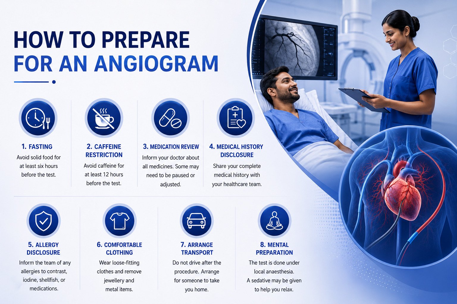
Fasting: Avoid solid food for at least six hours before the angiogram test. Clear liquids may be allowed up to two hours before, as directed by the cardiologist.
Caffeine restriction: Avoid caffeine for at least 12 hours before the test particularly before a CT coronary angiogram as caffeine raises heart rate and reduces the clarity of images.
Medication and blood thinners review: Inform the healthcare provider about all current medications. Blood thinners, diabetes medications, and certain supplements may need to be paused or adjusted hours before the test.
Medical history disclosure: The healthcare team will review the patient’s complete medical history, including any prior heart disease, kidney damage, high blood pressure, or high cholesterol.
Allergy disclosure: Inform the medical team of any known allergies to contrast agent, iodine, shellfish, or medications.
Comfortable clothing: Wear loose-fitting clothes and remove all jewellery and metal items before entering the cath lab.
Arrange transport: Patients must not drive after the procedure. Arrange for a family member or friend to accompany them home.
Mental preparation: The procedure is performed under local anaesthesia. A sedative may be given to help patients relax. Patients remain awake and can communicate with the healthcare team throughout.
Get an Accurate Cardiac Diagnosis — Book an Appointment
SRM Global Hospitals in Chennai provides advanced cardiac catheterisation, invasive coronary angiography, and CT coronary angiogram services led by experienced interventional cardiologists and supported by state-of-the-art cardiac imaging technology.
Expert-led coronary angiography performed by senior interventional cardiologists in a fully equipped cardiac catheterisation laboratory
Advanced CT coronary angiogram for non-invasive coronary CT angiography with high-resolution imaging and plaque characterisation
Same-session intervention angioplasty and stenting performed immediately when coronary angiography shows a significant blockage
Dedicated cardiac care team including interventional cardiologists, cardiac surgeons, imaging radiologists, and nephrologists
Fractional flow reserve (FFR) and intravascular ultrasound for functional assessment beyond standard coronary angiography findings
Comprehensive pre-procedure evaluation including medical history review, ECG, blood tests, and allergy assessment
Post-procedure monitoring and follow-up to prevent bleeding, protect kidney function, and ensure safe recovery
Multidisciplinary heart team following clinical practice guidelines for complex coronary artery disease management
FAQs on Angiogram
1. What Is an Angiogram and Why Is It Used as a Diagnostic Tool?
An angiogram is a diagnostic imaging procedure that uses contrast dye and x-ray images to visualise blood vessels and identify blockages, narrowed arteries, or coronary stenosis that restricts blood flow. Interventional cardiologists use the coronary angiogram as the gold standard diagnostic tool when a patient has chest pain, difficulty breathing, or abnormal non-invasive test results suggesting reduced blood flow to the heart muscle.
2. What Is the Difference Between a CT Coronary Angiogram and Conventional Coronary Angiography?
Conventional coronary angiography is an invasive procedure requiring a thin tube called a catheter inserted through the wrist or groin artery to inject contrast dye directly into the coronary arteries. A CT coronary angiogram or coronary CT angiography is non-invasive and uses a CT scanner with contrast dye through an IV to create detailed images without catheter insertion. The CT coronary angiogram can additionally assess plaque buildup in the artery walls, distinguishing between calcified and non-calcified plaque to assess heart attack risk, which conventional angiography cannot provide.
3. Is an Angiogram Painful?
The angiogram procedure is performed under local anaesthesia at the catheter site, so patients do not feel pain during catheter insertion. Patients may receive a sedative before the procedure to help them relax. A brief warm or flushing sensation when the contrast agent is injected is normal and temporary. Minor soreness at the catheter site is common afterwards and resolves within a few days.
4. How Long Does a Coronary Angiography Procedure Take?
A diagnostic coronary angiography procedure typically takes 30 to 60 minutes. After the procedure, most patients can go home within four to six hours. If angioplasty and stenting are performed during the same session to improve blood flow, the total time will be longer. Patients should plan for a half-day to full-day hospital visit including preparation, the procedure, and post-procedure monitoring.
5. What Are the Risks of a Coronary Angiogram?
Coronary angiography is a well-established invasive procedure with serious complications occurring in less than 1% of cases when performed by experienced cardiologists. Common minor side effects include bruising or soreness at the catheter site, temporary flushing from the contrast dye, and minor bleeding. Less common risks include an allergic reaction to contrast agent, kidney damage, blood clot forms during the procedure, or irregular heartbeats. Rare but serious complications can include heart attack, stroke, or damage to blood vessels. The risk of complications may be higher in patients with kidney disease or severe allergies.
6. How Soon Can I Return to Normal Activities After an Angiogram?
Most patients can return to normal activities, including work, within a few days after the angiogram. However, patients should consult their doctor before resuming normal activities. Patients are typically advised to rest and avoid strenuous activities for at least a week after the procedure. It is also common to feel tired after the procedure, and patients are encouraged to get plenty of sleep during their recovery period.
7. Can a Coronary Angiogram Detect All Types of Heart Blockages?
Coronary angiography is the most accurate diagnostic tool for detecting significant blockages within the coronary arteries and directly visualising reduced blood flow. However, it primarily shows the inner diameter of the artery lumen. A CT coronary angiogram can additionally show plaque buildup along the artery walls, distinguishing between calcified and non-calcified plaque. Fractional flow reserve (FFR) measurement and intravascular ultrasound may also be used alongside the coronary angiogram to fully assess coronary stenosis severity.
8. Who Should Avoid a Conventional Coronary Angiogram?
Patients with severe kidney disease, active bleeding disorders, or serious allergic reactions to contrast agent may not be suitable for conventional invasive coronary angiography. In such cases, cardiologists and nephrologists at SRM Global Hospitals evaluate alternative approaches such as a CT coronary angiogram or magnetic resonance angiography with adjusted protocols to protect kidney function and heart health.
9. What Happens If the Coronary Angiogram Shows a Blockage?
If coronary angiography shows a significant blockage causing reduced blood flow to the heart muscle, interventional cardiologists at SRM Global Hospitals discuss the findings immediately. Depending on the location, severity, and number of blocked coronary arteries, treatment options include angioplasty with stent placement in the same session to restore blood flow, scheduling a separate intervention, coronary artery bypass graft surgery, or optimising medical therapy with lifestyle changes and medications. Coronary angiography helps the healthcare team determine the most appropriate treatment path based on clinical practice guidelines and the individual patient’s condition.
